© 2017, GENASSIST, Inc.
By Keith S. Wexler, MBA, Maternal Fetal Medicine, Prenatal Diagnosis and Biotech/Life Sciences Consultant, GENASSIST, Inc.
Paul Wexler, M.D., F.A.C.O.G., Medical Director, GENASSIST, Inc.
Clinical Professor, Department of OB/GYN, University of Colorado Health Sciences Center
Clinical Professor, Division of Genetics/Dept. of Pediatrics, Univ. of Colorado/The Children’s Hospital
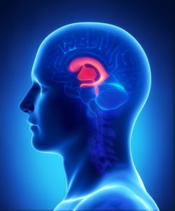
Choroid Plexus Cyst(s): A cyst or cysts found in the middle of the fetal brain which are small fluid filled spaces in the ventricles within the spongy layer of cells and vessels.
One or more of these cysts may be found on ultrasound in 1% to 2% of normal fetuses and 30% to 50% of fetuses with three #18 chromosomes (Trisomy 18 or Edward Syndrome).
Choroid Plexus Cyst(s) which can solitary or one of multiple ultrasound markers and can vary in size from 1 few millimeters to 25 millimeters or greater, is felt to be a strong ultrasound marker for Trisomy 18 and a possible indication for Maternal Fetal DNA, Chorionic Villus Sampling (CVS) or amniocentesis.
Choroid Plexus Cyst(s), like many isolated ultrasound findings should encourage the healthcare provider to perform a careful ultrasound evaluation to attempt to find other ultrasound markers suggestive of aneuploidy (an abnormal number of chromosomes).
Rarely, fetuses with Trisomy 21 (Down Syndrome) will also demonstrate a Choroid Plexus Cyst(s) but the relationship between Trisomy 21 and Choroid Plexus Cyst(s) is weak.
Ultrasound detection of a fetus with an increased likelihood of a trisomy is best for:
- Trisomy 13 (Patau Syndrome) – 90%
- Trisomy 18 (Edward Syndrome) – 80-90%
- Trisomy 21 (Down Syndrome) – 50% to 70%
Maternal Fetal DNA studies are best for detection of:
- Trisomy 21 (Down Syndrome) – Greater than 99%
- Trisomy 18 (Edward Syndrome) – 97% to 98%
- Trisomy 13 (Patau Syndrome) – 91% to 93%
*Chorionic Villus Sampling (CVS) and amniocentesis remain the best method for confirmation of an ultrasound marker for
Trisomy 13, Trisomy 18 or Trisomy 21. Chorionic Villus Sampling (CVS) is more likely to diagnose placental mosaicism and has a slightly greater miscarriage risk than amniocentesis, since the amniocentesis procedure is performed later in pregnancy.
Fetuses with Trisomy 18 often have several ultrasound markers in addition to the Choroid Plexus Cyst(s). However, we have followed two fetuses with Trisomy 18, confirmed by amniocentesis, that did not demonstrated any obvious ultrasound markers until after 18 weeks gestation and 21 weeks gestation respectively.
Ultrasound evaluation may identify additional Trisomy 18 markers including abnormal position of fingers, choroid plexus cyst(s), abnormal shaped head, 2 vessel cord, heart defects, intrauterine growth restriction (IUGR), omphalocele, neural tube defects and cystic hygroma.
As with all technology, a choroid plexus cyst(s) identified by ultrasound is not a confirmation that the baby has Trisomy 18.
Recommendation: **The patient needs to be aware of the guidelines from the American College of OB/GYN published in the OB/GYN Journal, Jan. 2007 recommends that “ideally, all women should be offered aneuploidy screening before 20 weeks gestation” and “all women regardless of age should have the option of invasive testing”.
