Tag Archives: #chromosomalabnormalities

Nuchal Translucency (NT) & Chromosomal Abnormalities in Pregnancy

© 2017, GENASSIST, Inc.     

By Keith S. Wexler, MBA, Maternal Fetal Medicine, Prenatal Diagnosis and Biotech /Life Science Consultant

Paul Wexler, M.D., F.A.C.O.G., Medical Director, GENASSIST, Inc.

Clinical Professor, Department of OB/GYN, University of Colorado Health Sciences Center

Clinical Professor, Division of Genetics/Dept. of Pediatrics, Univ. of Colorado/The Children’s Hospital 

Background: In the article “Nearly a Third of Abnormalities Found After 1st Trimester Screening Are Different Than Expected: 10 year Experience from a Single Center” by Alamillo, CM et al [in Prenatal Diagnosis 2013, 32, 1-6] results of 23,329 1st Trimester Screenings showed 6.3% were screen positive.

Using maternal age, nuchal translucency, free beta HCG and pregnancy associated plasma protein – A (PAPP-A) an abnormal karyotype was found in 3.9% of screen positive for Down Syndrome, 13.8% screen positives for Trisomy 13/18 and 45.9% of screen positives for Down Syndrome and Trisomy 13/18.

97 pregnancies were found to have an abnormal karyotype.  However:

  • 29.9% had chromosome abnormalities other than Trisomy 13, Trisomy 18 or Trisomy 21
  • 47,XXY and 45,X were the next most common chromosomal abnormalities found
  • There were 5 cases of mosaicism
  • 3 cases of polyploidy
  • 1 case each Trisomy 16 and Trisomy 22
  • 5 patients with structural chromosomal rearrangements

There was a direct relationship between the nuchal translucency thickness (NT) and the likelihood of a chromosomal abnormality.

  • NT between 3-5 mm & 3.9 mm, 10% of the fetuses had a chromosomal abnormality
  • NT between 4.0 mm & 4.9 mm, 40% of the fetuses had a chromosomal abnormality
  • NT between 5.0 mm & 5.9 mm, 46% of the fetuses had a chromosomal abnormality
  • NT between 6.0 and 6.9 mm, 50% of the fetuses had a chromosomal abnormality
  • NT between 7.0 mm & 7.9 mm, 40% of the fetuses had a chromosomal abnormality
  • NT greater than 8.2 mm, 100% (15 cases) had a chromosomal abnormality.

Although most healthcare providers would like to see Circulating Cell Free DNA (cfDNA) testing extended to all pregnant patients, the above data would suggest that:

1st Trimester Aneuploidy Screening is still a valuable tool for identifying fetuses at risk for other chromosomal abnormalities as well as for identifying structural abnormalities (e.g. gastroschisis, encephalocele, cystic hygroma, heart defects, limb abnormalities, diaphragmatic hernia and others).

Families should be counseled that circulating cell free fetal DNA testing although valuable, does not rule-out all chromosomal abnormalities and in the presence of abnormal 1st Trimester Screens or ultrasound findings more definitive testing by chorionic villus sampling (CVS) or amniocentesis may be indicated.

Analysis:

The availability of testing for Fetal DNA in the maternal circulation for Trisomy 13, Trisomy 18 and/or Trisomy 21 and the sex chromosomes [45,X/47,XXX/47,XXY and/or 47,XYY] for pregnant women at increased risk is a major breakthrough for those of us who have been awaiting a non-invasive test for chromosomal aneuploidy.

Combining the at risk population including advanced maternal age, family history of a chromosomal abnormality, abnormal ultrasound or biochemical tests, it is now possible to screen for some specific triploidies and sex chromosomal abnormalities.

However, we have known that Circulating Cell Free DNA (cf DNA) testing will need to evolve further to be able to diagnose chromosomal abnormalities other than the one’s described above and at present will miss most deletions, duplications, translocations and chromosomal mosaicism.

With the assistance of microarray testing and the advances in fetal DNA testing, the chromosomal abnormalities now missed will continue to be uncovered.

Choroid Plexus Cyst(s)

© 2017, GENASSIST, Inc.     

By Keith S. Wexler, MBA, Maternal Fetal Medicine, Prenatal Diagnosis and Biotech/Life Sciences Consultant, GENASSIST, Inc.

Paul Wexler, M.D., F.A.C.O.G., Medical Director, GENASSIST, Inc.

Clinical Professor, Department of OB/GYN, University of Colorado Health Sciences Center

Clinical Professor, Division of Genetics/Dept. of Pediatrics, Univ. of Colorado/The Children’s Hospital

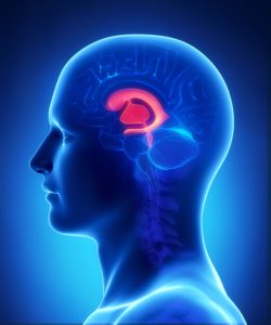
Choroid Plexus Cyst(s): A cyst or cysts found in the middle of the fetal brain which are small fluid filled spaces in the ventricles within the spongy layer of cells and vessels.

One or more of these cysts may be found on ultrasound in 1% to 2% of normal fetuses and 30% to 50% of fetuses with three #18 chromosomes (Trisomy 18 or Edward Syndrome). Choroid Plexus Cyst(s) which can solitary or one of multiple ultrasound markers and can vary in size from 1 few millimeters to 25 millimeters or greater, is felt to be a strong ultrasound marker for Trisomy 18 and a possible indication for Maternal Fetal DNA, Chorionic Villus Sampling (CVS) or amniocentesis.

Choroid Plexus Cyst(s), like many isolated ultrasound findings should encourage the healthcare provider to perform a careful ultrasound evaluation to attempt to find other ultrasound markers suggestive of aneuploidy (an abnormal number of chromosomes).

Rarely, fetuses with Trisomy 21 (Down Syndrome) will also demonstrate a Choroid Plexus Cyst(s) but the relationship between Trisomy 21 and Choroid Plexus Cyst(s) is weak.

Ultrasound detection of a fetus with an increased likelihood of a trisomy is best for:

  • Trisomy 13 (Patau Syndrome) – 90%
  • Trisomy 18 (Edward Syndrome) – 80-90%
  • Trisomy 21 (Down Syndrome) – 50% to 70%

Maternal Fetal DNA studies are best for detection of:

  • Trisomy 21 (Down Syndrome) – Greater than 99%
  • Trisomy 18 (Edward Syndrome) – 97% to 98%
  • Trisomy 13 (Patau Syndrome) – 91% to 93%

*Chorionic Villus Sampling (CVS) and amniocentesis remain the best method for confirmation of an ultrasound marker for

Trisomy 13, Trisomy 18 or Trisomy 21. Chorionic Villus Sampling (CVS) is more likely to diagnose placental mosaicism and has a slightly greater miscarriage risk than amniocentesis, since the amniocentesis procedure is performed later in pregnancy.

Fetuses with Trisomy 18 often have several ultrasound markers in addition to the Choroid Plexus Cyst(s). However, we have followed two fetuses with Trisomy 18, confirmed by amniocentesis, that did not demonstrated any obvious ultrasound markers until after 18 weeks gestation and 21 weeks gestation respectively.

Ultrasound evaluation may identify additional Trisomy 18 markers including abnormal position of fingers, choroid plexus cyst(s), abnormal shaped head, 2 vessel cord, heart defects, intrauterine growth restriction (IUGR), omphalocele, neural tube defects and cystic hygroma.

As with all technology, a choroid plexus cyst(s) identified by ultrasound is not a confirmation that the baby has Trisomy 18.

Recommendation: **The patient needs to be aware of the guidelines from the American College of OB/GYN published in the OB/GYN Journal, Jan. 2007 recommends that “ideally, all women should be offered aneuploidy screening before 20 weeks gestation” and “all women regardless of age should have the option of invasive testing”.

Ultrasound Markers – Choroid Plexus Cyst(s)

© 2017, GENASSIST, Inc.     

By Keith S. Wexler, MBA, Maternal Fetal Medicine, Prenatal Diagnosis and Biotech/Life Sciences Consultant, GENASSIST, Inc.

Paul Wexler, M.D., F.A.C.O.G., Medical Director, GENASSIST, Inc.

Clinical Professor, Department of OB/GYN, University of Colorado Health Sciences Center

Clinical Professor, Division of Genetics/Dept. of Pediatrics, Univ. of Colorado/The Children’s Hospital

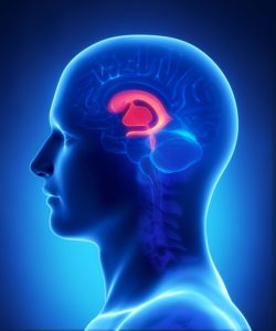
Choroid Plexus Cyst(s): A cyst or cysts found in the middle of the fetal brain which are small fluid filled spaces in the ventricles within the spongy layer of cells and vessels.

One or more of these cysts may be found on ultrasound in 1% to 2% of normal fetuses and 30% to 50% of fetuses with three #18 chromosomes (Trisomy 18 or Edward Syndrome).

Choroid Plexus Cyst(s) which can solitary or one of multiple ultrasound markers and can vary in size from 1 few millimeters to 25 millimeters or greater, is felt to be a strong ultrasound marker for Trisomy 18 and a possible indication for Maternal Fetal DNA, Chorionic Villus Sampling (CVS) or amniocentesis.

Choroid Plexus Cyst(s), like many isolated ultrasound findings should encourage the healthcare provider to perform a careful ultrasound evaluation to attempt to find other ultrasound markers suggestive of aneuploidy (an abnormal number of chromosomes).

Rarely, fetuses with Trisomy 21 (Down Syndrome) will also demonstrate a Choroid Plexus Cyst(s) but the relationship between Trisomy 21 and Choroid Plexus Cyst(s) is weak.

Ultrasound detection of a fetus with an increased likelihood of a trisomy is best for:

  • Trisomy 13 (Patau Syndrome) – 90%
  • Trisomy 18 (Edward Syndrome) – 80-90%
  • Trisomy 21 (Down Syndrome) – 50% to 70%

Maternal Fetal DNA studies are best for detection of:

  • Trisomy 21 (Down Syndrome) – Greater than 99%
  • Trisomy 18 (Edward Syndrome) – 97% to 98%
  • Trisomy 13 (Patau Syndrome) – 91% to 93%

*Chorionic Villus Sampling (CVS) and amniocentesis remain the best method for confirmation of an ultrasound marker for

Trisomy 13, Trisomy 18 or Trisomy 21. Chorionic Villus Sampling (CVS) is more likely to diagnose placental mosaicism and has a slightly greater miscarriage risk than amniocentesis, since the amniocentesis procedure is performed later in pregnancy.

Fetuses with Trisomy 18 often have several ultrasound markers in addition to the Choroid Plexus Cyst(s). However, we have followed two fetuses with Trisomy 18, confirmed by amniocentesis, that did not demonstrated any obvious ultrasound markers until after 18 weeks gestation and 21 weeks gestation respectively.

Ultrasound evaluation may identify additional Trisomy 18 markers including abnormal position of fingers, choroid plexus cyst(s), abnormal shaped head, 2 vessel cord, heart defects, intrauterine growth restriction (IUGR), omphalocele, neural tube defects and cystic hygroma.

As with all technology, a choroid plexus cyst(s) identified by ultrasound is not a confirmation that the baby has Trisomy 18.

Recommendation: **The patient needs to be aware of the guidelines from the American College of OB/GYN published in the OB/GYN Journal, Jan. 2007 recommends that “ideally, all women should be offered aneuploidy screening before 20 weeks gestation” and “all women regardless of age should have the option of invasive testing”.

Contact GENASSIST™

Fill out my online form.